Abdominal Anatomy | The lining of the uterus. The abdomen is the part of the body that contains all of the structures between the thorax (chest) and the pelvis, and is separated from the thorax via the diaphragm. The major muscles of the abdomen include the rectus abdominis in front, the external obliques at the sides, and the latissimus dorsi muscles in the back. The majority of these organs are encased in a protective membrane termed the peritoneum. Radiographers suggest an abdominal ct scan to look … atlas of ct anatomy of the abdomen read more »
These two apertures, together with abdominal walls, bound the abdominal cavity. 3d interactive models and tutorials on the anatomy of the abdomen and pelvis. Abdominal muscle, any of the muscles of the anterolateral walls of the abdominal cavity, composed of three flat muscular sheets, from without inward: The major muscles of the abdomen include the rectus abdominis in front, the external obliques at the sides, and the latissimus dorsi muscles in the back. Abdominal exercises 12 photos of the abdominal exercises ab exercises knee injury, abdominal exercises images, abdominal exercises on the total gym, abdominal exercises with hernia, abdominal exercises yoga, human anatomy, ab exercises knee injury, abdominal exercises images, abdominal exercises on the total gym, abdominal exercises with.
It is the long, flat muscle that extends vertically between the pubis and the fifth, sixth, and seventh ribs. Inferiorly the abdomen is open to the pelvis, communicating through the superior pelvic aperture (pelvic inlet). The venous drainage of the abdomen is primarily mediated through the portal venous system and the inferior vena cava (ivc). The anterolateral abdominal wall consists of four main layers (external to internal): Anatomy of female pelvic area. The major muscles of the abdomen include the rectus abdominis in front, the external obliques at the sides, and the latissimus dorsi muscles in the back. External oblique, internal oblique, and transverse abdominis, supplemented in front on each side of the midline by rectus abdominis. For the sake of brevity, the various organs will be not discussed in detail. The major organs of the abdomen include the. Abdominal computed tomography abdominal computed tomography (ct) is a type of medical imaging procedure used to diagnose and monitor internal stomach issues, like cancer, bowel obstruction, and abdominal pain. The diaphragm is its upper boundary. The abdomen is the front part of the abdominal segment of the trunk. These layers are a bit different between the umbilical region and the groin, but overall the basic layers are the same.
The rectus abdominis connects to the xiphoid process, a bony landmark at the bottom of the sternum. Labeled scrollable ct of the abdomen covering anatomy with a level of detail appropriate for medical students. The major organs of the abdomen include the. The normal anatomy or organs imaged in a standard abdominal examination is explained below. The diaphragm forms the upper surface of the abdomen.
The area occupied by the abdomen is called the abdominal cavity. Labeled scrollable ct of the abdomen covering anatomy with a level of detail appropriate for medical students. The abdominal wall is subdivided into the anterior wall, the right and left lateral walls, and the posterior wall. The region occupied by the abdomen is called the abdominal cavity, and is enclosed by the abdominal muscles at front and to the sides, and by part of the vertebral column at the back. An abdominal aortic aneurysm consists of a weakening of the wall of the aorta just above the point where it bifurcates into the left and right common iliac arteries. The abdomen (colloquially called the stomach, belly, tummy or midriff) is the part of the body between the thorax (chest) and pelvis, in humans and in other vertebrates. The rectus abdominis connects to the xiphoid process, a bony landmark at the bottom of the sternum. Chapter 76 venous anatomy of the abdomen and pelvis. The abdomen is the body region found between the thorax and the pelvis. The lining of the uterus. 3d interactive models and tutorials on the anatomy of the abdomen and pelvis. Abdominal exercises 12 photos of the abdominal exercises ab exercises knee injury, abdominal exercises images, abdominal exercises on the total gym, abdominal exercises with hernia, abdominal exercises yoga, human anatomy, ab exercises knee injury, abdominal exercises images, abdominal exercises on the total gym, abdominal exercises with. Abdominal muscle, any of the muscles of the anterolateral walls of the abdominal cavity, composed of three flat muscular sheets, from without inward:
Two female reproductive organs located in the pelvis. Abdomen, in human anatomy, the body cavity lying between the chest or thorax above and the pelvis below and from the spine in the back to the wall of abdominal muscles in the front. The abdomen is the part of the body that contains all of the structures between the thorax (chest) and the pelvis, and is separated from the thorax via the diaphragm. 3d interactive models and tutorials on the anatomy of the abdomen and pelvis. An abdominal aortic aneurysm consists of a weakening of the wall of the aorta just above the point where it bifurcates into the left and right common iliac arteries.

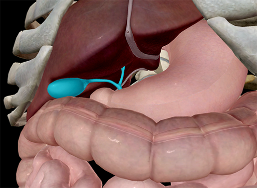
These layers are a bit different between the umbilical region and the groin, but overall the basic layers are the same. It is divided into the fundus, body, antrum and pylorus, and its blood supply is derived from the coeliac trunk. The abdomen is the part of the body that contains all of the structures between the thorax (chest) and the pelvis, and is separated from the thorax via the diaphragm. From the outside to the inside is the skin, then a layer of fat. The abdomen (commonly called the belly) is the body space between the thorax (chest) and pelvis. Abdominal computed tomography (ct) is a type of medical imaging procedure used to diagnose and monitor internal stomach issues, like cancer, bowel obstruction, and abdominal pain. The rectus abdominis connects to the xiphoid process, a bony landmark at the bottom of the sternum. The major muscles of the abdomen include the rectus abdominis in front, the external obliques at the sides, and the latissimus dorsi muscles in the back. Chapter 76 venous anatomy of the abdomen and pelvis. The region occupied by the abdomen is called the abdominal cavity, and is enclosed by the abdominal muscles at front and to the sides, and by part of the vertebral column at the back. The venous drainage of the abdomen is primarily mediated through the portal venous system and the inferior vena cava (ivc). Anatomy of female pelvic area. Abdominal exercises 12 photos of the abdominal exercises ab exercises knee injury, abdominal exercises images, abdominal exercises on the total gym, abdominal exercises with hernia, abdominal exercises yoga, human anatomy, ab exercises knee injury, abdominal exercises images, abdominal exercises on the total gym, abdominal exercises with.
Abdominal Anatomy: In anatomy and physiology, you'll learn how to divide the abdomen into nine different regions and four different quadrants.
Refference: Abdominal Anatomy
Abdominal Anatomy | The lining of the uterus. The abdomen is the part of the body that contains all of the structures between the thorax (chest) and the pelvis, and is separated from the thorax via the diaphragm. The major muscles of the abdomen include the rectus abdominis in front, the external obliques at the sides, and the latissimus dorsi muscles in the back. The majority of these organs are encased in a protective membrane termed the peritoneum. Radiographers suggest an abdominal ct scan to look … atlas of ct anatomy of the abdomen read more »
These two apertures, together with abdominal walls, bound the abdominal cavity. 3d interactive models and tutorials on the anatomy of the abdomen and pelvis. Abdominal muscle, any of the muscles of the anterolateral walls of the abdominal cavity, composed of three flat muscular sheets, from without inward: The major muscles of the abdomen include the rectus abdominis in front, the external obliques at the sides, and the latissimus dorsi muscles in the back. Abdominal exercises 12 photos of the abdominal exercises ab exercises knee injury, abdominal exercises images, abdominal exercises on the total gym, abdominal exercises with hernia, abdominal exercises yoga, human anatomy, ab exercises knee injury, abdominal exercises images, abdominal exercises on the total gym, abdominal exercises with.
It is the long, flat muscle that extends vertically between the pubis and the fifth, sixth, and seventh ribs. Inferiorly the abdomen is open to the pelvis, communicating through the superior pelvic aperture (pelvic inlet). The venous drainage of the abdomen is primarily mediated through the portal venous system and the inferior vena cava (ivc). The anterolateral abdominal wall consists of four main layers (external to internal): Anatomy of female pelvic area. The major muscles of the abdomen include the rectus abdominis in front, the external obliques at the sides, and the latissimus dorsi muscles in the back. External oblique, internal oblique, and transverse abdominis, supplemented in front on each side of the midline by rectus abdominis. For the sake of brevity, the various organs will be not discussed in detail. The major organs of the abdomen include the. Abdominal computed tomography abdominal computed tomography (ct) is a type of medical imaging procedure used to diagnose and monitor internal stomach issues, like cancer, bowel obstruction, and abdominal pain. The diaphragm is its upper boundary. The abdomen is the front part of the abdominal segment of the trunk. These layers are a bit different between the umbilical region and the groin, but overall the basic layers are the same.
The rectus abdominis connects to the xiphoid process, a bony landmark at the bottom of the sternum. Labeled scrollable ct of the abdomen covering anatomy with a level of detail appropriate for medical students. The major organs of the abdomen include the. The normal anatomy or organs imaged in a standard abdominal examination is explained below. The diaphragm forms the upper surface of the abdomen.
The area occupied by the abdomen is called the abdominal cavity. Labeled scrollable ct of the abdomen covering anatomy with a level of detail appropriate for medical students. The abdominal wall is subdivided into the anterior wall, the right and left lateral walls, and the posterior wall. The region occupied by the abdomen is called the abdominal cavity, and is enclosed by the abdominal muscles at front and to the sides, and by part of the vertebral column at the back. An abdominal aortic aneurysm consists of a weakening of the wall of the aorta just above the point where it bifurcates into the left and right common iliac arteries. The abdomen (colloquially called the stomach, belly, tummy or midriff) is the part of the body between the thorax (chest) and pelvis, in humans and in other vertebrates. The rectus abdominis connects to the xiphoid process, a bony landmark at the bottom of the sternum. Chapter 76 venous anatomy of the abdomen and pelvis. The abdomen is the body region found between the thorax and the pelvis. The lining of the uterus. 3d interactive models and tutorials on the anatomy of the abdomen and pelvis. Abdominal exercises 12 photos of the abdominal exercises ab exercises knee injury, abdominal exercises images, abdominal exercises on the total gym, abdominal exercises with hernia, abdominal exercises yoga, human anatomy, ab exercises knee injury, abdominal exercises images, abdominal exercises on the total gym, abdominal exercises with. Abdominal muscle, any of the muscles of the anterolateral walls of the abdominal cavity, composed of three flat muscular sheets, from without inward:
Two female reproductive organs located in the pelvis. Abdomen, in human anatomy, the body cavity lying between the chest or thorax above and the pelvis below and from the spine in the back to the wall of abdominal muscles in the front. The abdomen is the part of the body that contains all of the structures between the thorax (chest) and the pelvis, and is separated from the thorax via the diaphragm. 3d interactive models and tutorials on the anatomy of the abdomen and pelvis. An abdominal aortic aneurysm consists of a weakening of the wall of the aorta just above the point where it bifurcates into the left and right common iliac arteries.
These layers are a bit different between the umbilical region and the groin, but overall the basic layers are the same. It is divided into the fundus, body, antrum and pylorus, and its blood supply is derived from the coeliac trunk. The abdomen is the part of the body that contains all of the structures between the thorax (chest) and the pelvis, and is separated from the thorax via the diaphragm. From the outside to the inside is the skin, then a layer of fat. The abdomen (commonly called the belly) is the body space between the thorax (chest) and pelvis. Abdominal computed tomography (ct) is a type of medical imaging procedure used to diagnose and monitor internal stomach issues, like cancer, bowel obstruction, and abdominal pain. The rectus abdominis connects to the xiphoid process, a bony landmark at the bottom of the sternum. The major muscles of the abdomen include the rectus abdominis in front, the external obliques at the sides, and the latissimus dorsi muscles in the back. Chapter 76 venous anatomy of the abdomen and pelvis. The region occupied by the abdomen is called the abdominal cavity, and is enclosed by the abdominal muscles at front and to the sides, and by part of the vertebral column at the back. The venous drainage of the abdomen is primarily mediated through the portal venous system and the inferior vena cava (ivc). Anatomy of female pelvic area. Abdominal exercises 12 photos of the abdominal exercises ab exercises knee injury, abdominal exercises images, abdominal exercises on the total gym, abdominal exercises with hernia, abdominal exercises yoga, human anatomy, ab exercises knee injury, abdominal exercises images, abdominal exercises on the total gym, abdominal exercises with.
Abdominal Anatomy: In anatomy and physiology, you'll learn how to divide the abdomen into nine different regions and four different quadrants.
Refference: Abdominal Anatomy
Show Disqus Comment Hide Disqus Comment